CURRENT PROJECTS
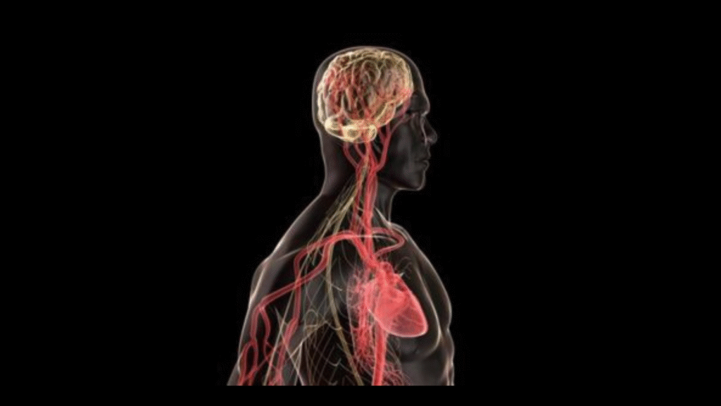
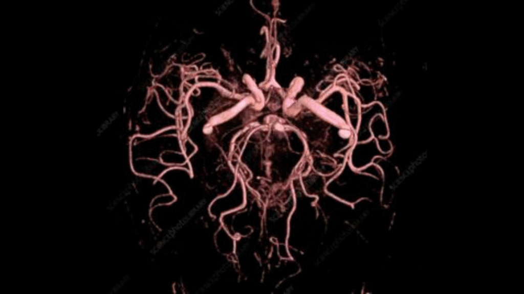
Acute Stroke Imaging
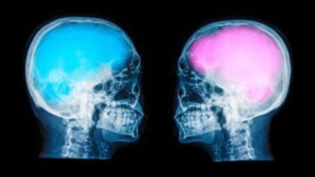
Sex Differences in Acute Stroke
A growing body of literature indicates that the severity of a stroke, response to treatment, and functional outcomes following stroke differ by sex. Dr. Dula’s primary research initiative involves examining the role of anatomical and hemodynamic (bloodflow dynamics) factors in observed sex differences in stroke. Our current study—Women’s Imaging of Stroke Hemodynamics Study (WISHeS)—aims to identify factors which differentiate people by sex in terms of their stroke symptoms, the severity and type of their stroke, response to different types of treatment, and their functional recovery.
Both retrospective data and prospective data collected specifically for WISHeS is being analyzed as part of this initiative, in collaboration with the Lone Star Stroke Research Consortium as well as collaborators at UT Houston, Stanford, and UCLA.
Funding: Career Development Award from the American Heart Association, Lone Star Stroke Research Consortium
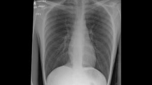
Code Stroke Imaging (CSI) Austin: MRI or CT?
When someone with a suspected stroke arrives at the hospital, one of the first and most crucial tasks in determining a diagnosis and potential treatment options is obtaining imaging of the brain. Currently, stroke patients first have their brain imaged either using Magnetic Resonance (MR) or a Computed Tomography (CT) scanner, depending on availability of equipment, personnel, and other practical considerations. However, there may be benefits to prioritizing one modality over another. For instance, MR imaging can show more detailed images of the brain, helping to better localize the area where a stroke occurred and better informing treatment options, while CT imaging can be completed much more quickly—an important consideration when time is crucial. Our pilot study intends to determine the feasibility of randomizing the assignment of first imaging modality (i.e., MRI or CT) to incoming suspected stroke cases. This is the first step in developing a larger international study in which the benefits of using MRI or CT first can be examined related to functional and treatment outcomes, potentially leading to improved standards of care across US and international hospitals.
Funding: Lone Star Stroke Research Consortium
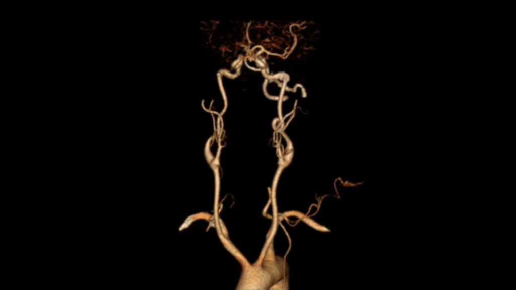
 Magnetic Resonance Imaging Predictors of Atrial Fibrillation in Cryptogenic Stroke Cases
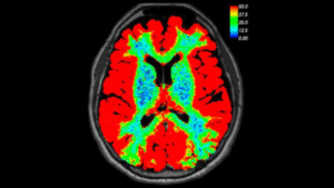
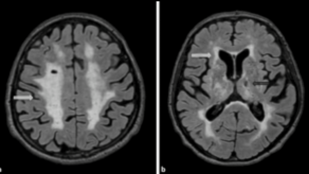
Understanding the origin of an ischemic stroke is key in both providing effective treatment and in implementing protective measures to avoid recurrence of stroke. Approximately 25% of ischemic strokes are determined to be embolic strokes of unknown source, where it is difficult to determine whether or not the source is cardioembolic (related to an embolus within the heart). Methods such as administration of anticoagulants or long-term cardiac monitoring can be employed in order to help prevent stroke recurrence when the source of the stroke is defined as cardioembolic. It is theorized that a definable imaging pattern with lesions in multiple vascular territories (MVT; multiple areas in the brain) correlates to strokes of cardioembolic origin. This study aims to develop and validate methods for imaging and identifying MVT in order to more accurately identify strokes with likely cardioembolic sources where preventative measures can be implemented to avoid another stroke. Implications of validating this method of identifying an acute brain imaging biomarker include a reduction in secondary stroke rate, death, disability, and healthcare costs.
Funding: Lone Star Stroke Research Consortium
Glymphatics
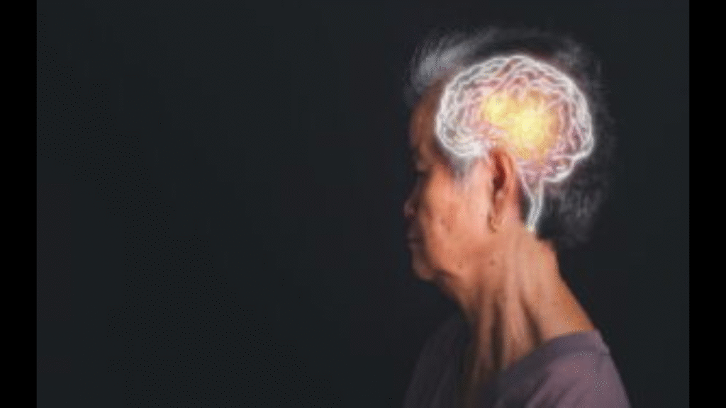
Laterality of Glymphatics in Parkinson’s Disease
In the initial stages of Parkinson’s disease (PD), symptoms are isolated to one side of the body for a period of time. While the mechanism underlying this is currently unknown, we suspect that the functioning of the glymphatic system, which helps to clear waste in the brain during sleep, may play a role. In our study on glymphatics in PD, we will validate a new application of MR imaging techniques for recording active functioning of the glymphatic system. Through analysis of the imaging, we hope to illuminate the role of waste clearance in the pathophysiology of Parkinson’s disease. Results from this study have the potential to inform development of targeted immunomodulatory therapeutics that could restore brain-immune system communication, support plasticity and repair, and modify the course of neurodegeneration. Additionally, the validated MR imaging methods for glymphatics could aid more broadly in allowing researchers to explore the role of the glymphatic system in other neurodegenerative diseases and healthy aging.
Funding: UT Cain Foundation Pilot Award
Open and Accessible Clinical Imaging
Accessible Imaging Data for Clinical Trials
Clinical research trials largely still employ burdensome methods for data collection, organization, and study operations. Medical records and reports needed for data collection are printed (sometimes for a fee) and stored on paper, and medical images are transferred to CDs only accessible with proprietary software and sent through the mail. These methods are inefficient and costly for patients, trial sites, and clinical data coordinating centers, and slow the progress and translation of evidence-based improvements in medical care. Yet, 85% of Americans have the ultimate data capture tool in their pockets—the smartphone. Our research goal is to establish a cost-effective, safe method for capture and transmit medical imaging via smartphone technology, aiding in efficiency of clinical research and allowing for improved access and service for underserved patient populations.
Funding: UT Health Catalyst
Automatic Extraction of Commercial Image Processing Output
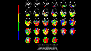
Commercial software is available and widely used in the clinic to obtain stroke lesion characteristics and determine treatment eligibility. These platforms are extremely useful in the clinic, however, the data output are nonconventional and difficult to use for research. As we did not have access to the intermediate results of the processing done by the software used to produce these images, our objective was to obtain the data from the summary images themselves. Therefore, we used Optical Character Recognition (OCR) to extract the textual information from each of the summary images. We used optical character recognition (OCR) to extract the textual information from the summary images resulting from the commercial software found on the hospital imaging system.
As we were unable to improve OCR results significantly via preprocessing or altering hyperparameters, we opted to perform error correction on the extracted text. Due to domain-specific terminology and scientific structuring of the data we opted for a customized approach rather than using a general error correction model for the English language. Because many of the errors were small perturbations to the actual data, we implemented a novel error correction algorithm that combines dictionary-based correction with iterative randomized character substitutions of very frequent errors. This approach was able to accurately correct all the OCR outputs from all images (verified with manual review), which could then be parsed with regex and used to construct a dataframe. Randomized substitution of frequently occurring misclassifications is especially useful in addressing the stochastic nature of spacing-related issues in OCR that are difficult to resolve purely with dictionary-based correction. Applying this iteratively until a stable outcome is reached increases the likelihood of correcting OCR misclassifications that critically impair dictionary-based correction. The novel algorithm allowed extraction of stroke lesion volume information for further quantification of these legacy data.
Repository / Open Science Contributions
 ENIGMA-Stroke Recovery
ENIGMA-Stroke Recovery is part of the ENIGMA Consortium, an international effort to understand brain structure and function through researchers in imaging genomics, neurology and psychiatry. ENIGMA-Stroke Recovery is a working group dedicated to improving our understanding of how changes in the brain after stroke relate to functional outcomes and recovery. This group is actively conducting meta- and mega-analyses of existing MRI data in adults who have had a stroke. The ImgARC Lab routinely contributes data and is currently doing a secondary analysis to evaluate sex differences in stroke using the ENIGMA-Stroke Recovery database.
A Multi-Institutional Chest Radiograph Dataset To Evaluate AI Generalizability (MAIDA)
Led by researchers at Harvard, a large multi-institutional international effort is underway to build a dataset of chest radiographs to train and evaluate AI interpretation models. Ultimately, these models could have applications aiding in diagnostic processes in various clinical settings. The creation of a public repository of chest imaging data collected from a range of clinical settings and populations internationally will enable exploration of the extent and limit of generalizability of machine learning models with an eye toward equity and safety. The consortium of participating hospitals is expanding rapidly, and we are continually looking for site PIs interested in responsible applications of AI models in clinical settings to join.

Medical Imaging and Data Resource Center (MIDRC) Data contribution
The ImgARC Lab is helping to contribute data to a national repository of images and other clinical data related to COVID-19 in order to fuel studies advancing our understanding of how to diagnose, monitor, and treat the virus. Imaging and clinical data related to COVID-19 is gathered, deidentified, and uploaded to the repository for use in research.
Funding: RSNA MIDRC Award
Cerebrovascular Health and Brain Age
Coronary Artery Risk Development in Young Adults (CARDIA) Brain MRI Study
The Coronary Artery Risk Development in Young Adults (CARDIA) study is a large-scale longitudinal study that examines the development of and predictors of cardiovascular disease. Longitudinal data collected include a wealth of clinical data as well as functional brain imaging data collected from a subset of participants at three study timepoints. The ImgARC Lab is a brain MRI processing site for fMRI and cerebrovascular reactivity data for the CARDIA study. Additionally, we are leading a study on brain aging leveraging data from CARDIA to determine whether cardiovascular risk factors are associated with functional brain changes and whether this association varies by sex and race.
Funding: Subaward from University of Pennsylvania via NIH
CARDIA – Cerebrovascular Brain Aging
Early identification of risk of vascular dysfunction in middle-aged individuals has the potential to help avoid medical emergencies, aid in mitigating symptoms, and reduce cognitive and social effects of cerebrovascular disease. Current machine learning-based models aimed at early identification of cerebrovascular dysfunction have relied on structural outcome measures such as measures of brain atrophy. Using machine learning methods, we will develop a novel brain age prediction model derived from measures of cerebrovascular function, which may precede observable structural abnormalities. We will characterize the spectrum of age-related microvascular, functional, and phenotypic changes across normal aging populations, individuals with subclinical vascular dysfunction, and overt cerebrovascular disease. With the aggregation of a large dataset and evaluation of sex as a biological variable, we hope to ensure stability and generalizability of a model with prognostic utility to predict overt cerebrovascular disease and cognitive decline.
Funding: Subaward from University of Pennsylvania via NIH
Recognizing Stroke in Women
Stroke poses a significant threat, often leading to severe disability or even death. Timely recognition of stroke symptoms is key in mitigating stroke symptoms, increasing a person’s chances of effective treatment and recovery. Enhancing public knowledge about stroke is pivotal to designing effective preventive measures to combat disabling symptoms and premature mortality. The ImgARC Lab participates as a research site for the Women and Stroke, Knowledge, Awareness, and Confidence Level Survey (WASKACS), an initiative to assess the general public’s understanding of sex-specific stroke risks and symptoms in females. This educational survey—available in English and Spanish—will inform initiatives for improving education and preparedness surrounding stroke.
COVID Brain Perfusion
Assess Cerebral Blood Flow in Long COVID Patients
This project assesses cerebral blood flow in long COVID patients as well as examine the neural correlates of cognitive dysfunction after COVID-19. We expect that survivors of mild to moderate COVID-19 will demonstrate significantly lower executive-attention network connectivity compared to non-infected controls and that this will correlate with cognitive dysfunction. We are specifically focusing on mechanism underlying cognitive impairment in COVID-19 associated with cerebral perfusion
This work is in collaboration with Dr. Shelli Kesler
Placental Perfusion and Outcomes
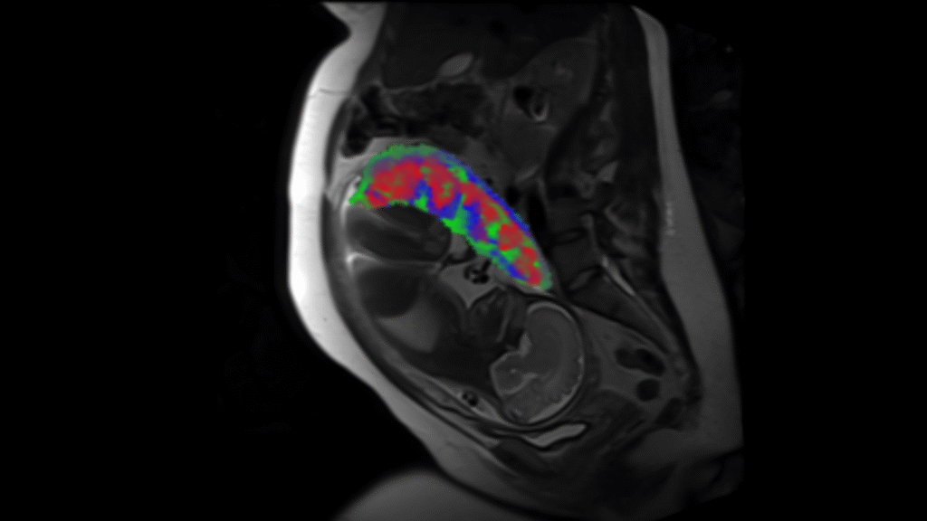
Perfusion Measures in Pregnancy Associated with Nutrition & Fetal Outcomes
Maternal obesity, dietary intake, and nutrition status during pregnancy have been linked with preeclampsia, gestational hypertension, maternal cardiovascular biomarkers during pregnancy (insulin, insulin resistance and triglycerides), and child birth outcomes in epidemiologic research. The placenta is likely enmeshed in the causal pathways, but we have limited understanding of how these exposuresimpact placentation and placental function. Abnormal placentation and associated consequences are theorized to impact the pathophysiology of preeclampsia and other adverse outcomes, and nutritional status and dietary intake may be a key potentially modifiable factor in these associations.
We are assessing perfusion measures in the placenta associated with nutrition and fetal outcomes. Specifically, we are looking at microcirculation within different layers of the placenta to evaluate early markers of maternal or fetal health.
This work is in collaboration with Dr. Beth Widen
PREVIOUS PROJECTS
Tenecteplase As Stroke Thrombolytic in Clinical Practice
Prospective Observational Cohort Study of Tenecteplase As Stroke Thrombolytic in Clinical Practice
Alteplase is currently the only thrombolytic approved by US Food and Drug Administration (FDA) for the treatment of ischemic stroke. Tenecteplase, FDA approved only for use in ST-elevation myocardial infarction, is given as a 5-10 second single bolus, as opposed to bolus plus 60-minute infusion for alteplase, owing to greater fibrin specificity and a longer half-life than alteplase. We performed a prospective registry-based observational, sequential cohort comparison tenecteplase (n=234) to alteplase (n=354) treated stroke patients. We found that switching to tenecteplase in routine clinical practice in a 10-hospital network was associated with shorter DTN and DIDO times, non-inferior favorable clinical outcomes at discharge, and reduced hospital costs. Evaluation in larger, multicenter cohorts is recommended to determine if these observations generalize.