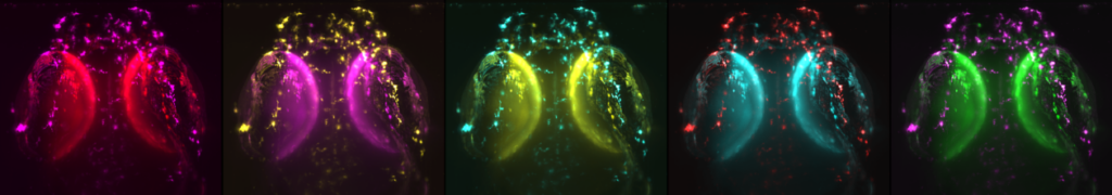
Our main research focus is to understand the molecular signals that cue regeneration of the retinal pigment epithelium (RPE). The RPE is a monolayer of cells located at the back of the eye at a critical interface between the retinal photoreceptors and an ocular blood supply called the choroidal vasculature. The RPE performs a myriad of functions important for maintaining retinal health and vision, and loss of RPE tissue leads to visual impairment or blindness. We use the zebrafish as a model system to study how the RPE regenerates after injury and we are specifically interested in how the immune response regulates this regenerative response.
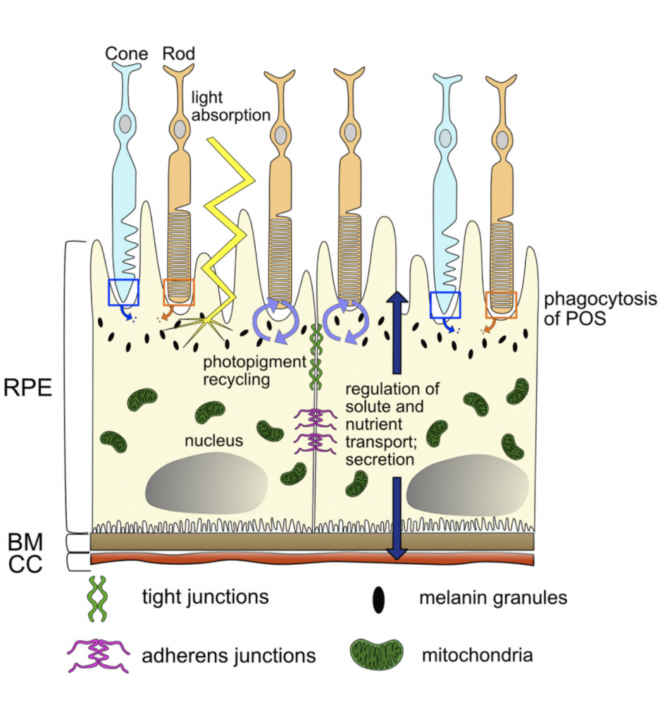
RPE biology and generalized functions (above left). Cartoon illustrating the mature, polarized RPE monolayer and its interactions with the rod and cone PRs, BM, and CC, as well as the diversity of functions mediated by the RPE. Abbreviations: PRs, photoreceptors; RPE, retinal pigment epithelium; CC, choriocapillaris; BM, Bruch’s membrane; POS, photoreceptor outer segments. Schematic and text from Figure 1 in George et al., PRER 2021.
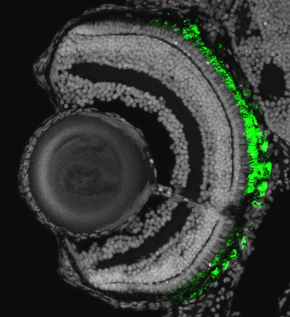
Larval zebrafish eye (above right). Fluorescent cross-section image showing the tissues of a 5-day-old zebrafish eye, including the lens (left), 3 cellular layers of the neural retina, and the RPE (green). In this transgenic zebrafish line (rpe65a:nfsB-eGFP), the RPE is specifically labeled with green fluorescent protein (eGFP) for visualization and also expresses nitroreductase (NTR/nfsB) for injury induction. White labels all cell nuclei; dorsal is up, ventral is down.