• Skip to primary navigation
  • Skip to main content
  • Skip to footer
UT Shield
The University of Texas at Austin
  • Home
  • About
  • Research
  • News
  • Publications
  • Lab Members

Research

August 15, 2025, Filed Under: Research

Self-Assembling Peptides

  • Design of self-assembling, peptide-based hydrogels for drug delivery and tissue engineering

Summary: Scaffolds play essential roles in drug delivery and tissue engineering. They not only provide the architecture to deliver therapeutics and to support cells, but also physical cues for cell behavior, including attachment, proliferation, and gene expression. Superior scaffolds have a variety of characteristics in common: they have a physical structure similar to the extracellular matrix (ECM); they are robust enough to support either drug loading and controlled release or cell attachment and cellular expansion; they are highly hydrated at physiological conditions; they are diffusive enough to allow nutrients, growth factors, or bioactive molecules to reach cells in addition to facilitating the removal of cellular wastes; and they have built-in degradability that allows for easy clearance with nontoxic byproducts.

Protein-inspired, peptide hydrogels are promising for biomaterials applications. Hydrogels made from self-assembling peptides have significant potential in drug delivery and tissue engineering because they provide a highly hydrated, ECM-like environment with inherent biocompatibility and straightforward tunability due to the nature and character of amino acids; such constructs therefore have characteristics that mimic the natural environment and can be readily designed for specific purposes. Small molecule, self-assembling peptides are a promising new field in scaffold-design research.


Hydrogels for 3D Cell Cultures

Project One
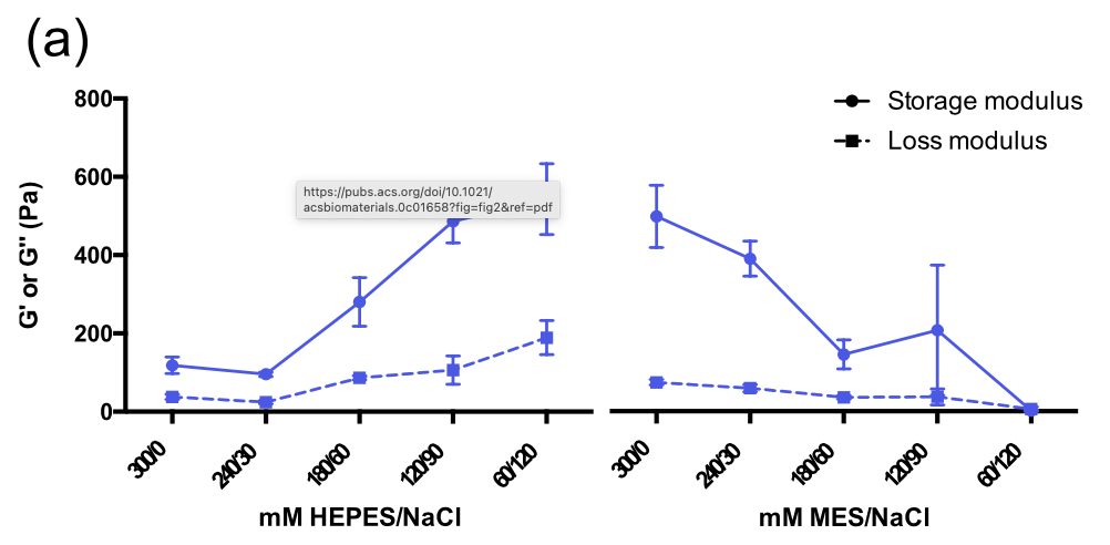
We are investigating novel nucleopeptide hydrogel scaffolds to mimic a three-dimensional (3D) environment. Current 3D hydrogels for cell culture, like Matrigel, are widely used but inherently flawed due to their biological origin. By constructing a synthetic peptide sequence which undergoes gelation in physiological conditions we aim to develop a system that is modular and reproducible. Herein, we have assesed numerous combinations of short amino acid sequences to create a hydrogel capable of supporting cells in a 3D environment. Diverging from traditional solid-phase peptide synthesis, we have substituted the controversial Fmoc molecule with biocompatible nucleobases.


Carrier for miRNA cargo

Project Two

We further aim to capitalize on our unique nucleopeptides by analyzing their capacity to act as a vector for nucelic acids, specifically microRNA (miRNA). Our material will inherently intercalate with the cargo to facilitate entry into cells and improve genetic uptake by overcoming at least one of the biological barriers associated with therapeutic delivery.

August 15, 2025, Filed Under: Research

Extracellular Matrix Stiffness

  • Dynamic ECM stiffening alters cell phenotype and tumor progression

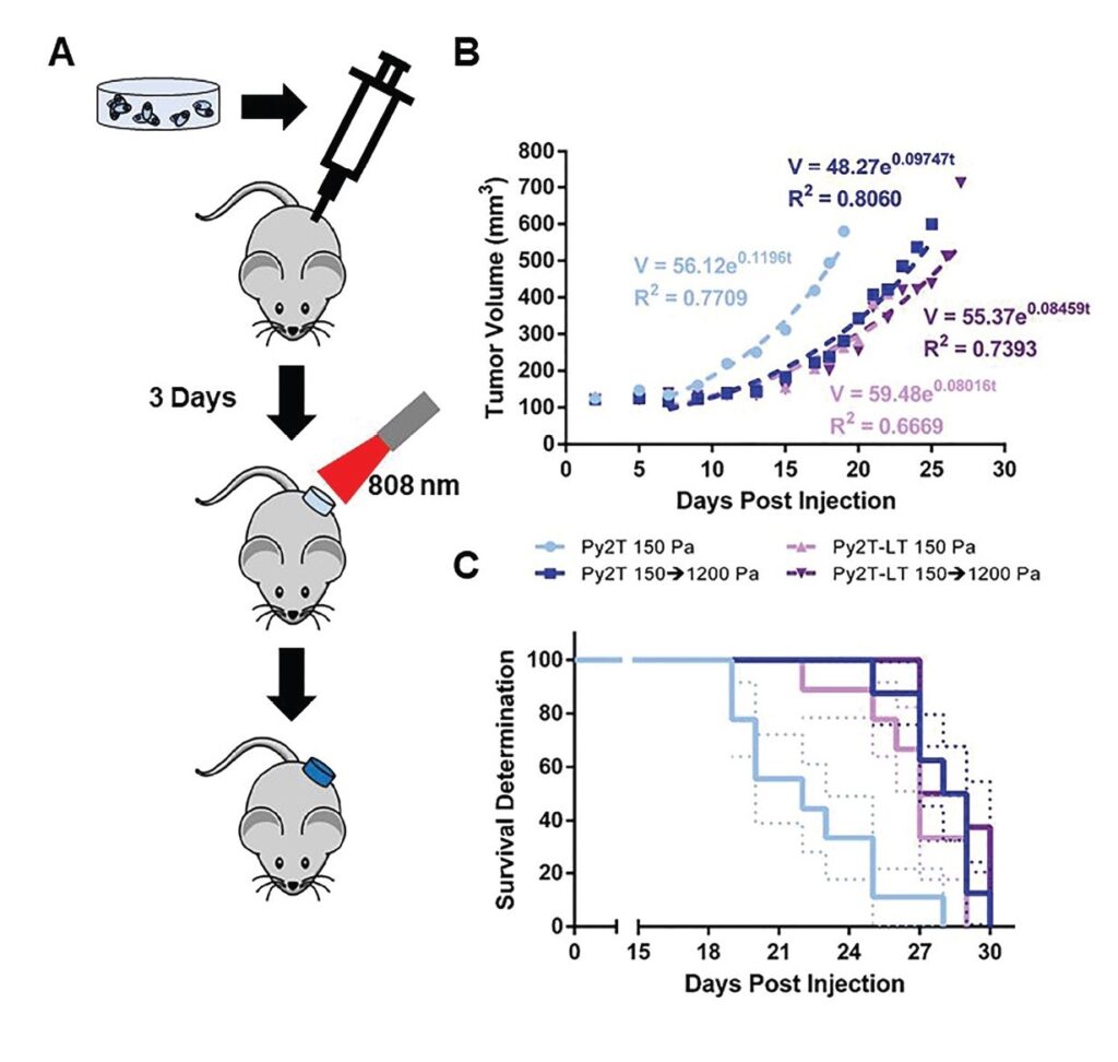
Summary: Soft tissue tumors, including breast cancer, become stiffer throughout disease progression. This increase in stiffness has been shown to correlate to malignant phenotype and epithelial-to-mesenchymal transition (EMT) in vitro. Unlike current models, utilizing static increases in matrix stiffness, our group has previously created a system that allows for dynamic stiffening of an alginate–matrigel composite hydrogel to mirror the native dynamic process. Here, we utilize this system to evaluate the role of matrix stiffness on EMT and metastasis both in vitro and in vivo. Epithelial cells were seen to lose normal morphology and become protrusive and migratory after stiffening. This shift corresponded to a loss of epithelial markers and gain of mesenchymal markers in both the cell clusters and migrated cells. Furthermore, stiffening in a murine model reduced tumor burden and increased migratory behavior prior to tumor formation. 

August 15, 2025, Filed Under: Research

Macrophage Repolarization

  • Characterizing macrophage repolarization by engulfed apoptotic cells in regenerative medicine

Summary: Macrophages that have engulfed apoptotic cells tend to polarize toward an anti-inflammatory (M2) phenotype over a pro-inflammatory (M1) phenotype. Our lab is interested in which characteristics of apoptotic cells cause this shift. We also utilize the anti-inflammatory nature of apoptotic cells to produce nanoparticle systems to aid in the calming of inflammation for regenerative medicine.


Engineering Nanoparticles to Control Macrophage Phenotype

Project One

Current anti-inflammatory drugs have significant drawbacks, including variable response rates and off-target effects. Here, we have developed an apoptotic-body inspired nanoparticle to modulate inflammatory macrophage phenotype. This polymeric nanoparticle is coated with phosphatidylserine-supplemented cell plasma membrane to mimic the anti-inflammatory effect of apoptotic cell engulfment. Nanoparticle delivery reduces inflammatory cytokine production and promotes an anti-inflammatory phenotypic macrophage shift. The capacity of these nanoparticles to help resolve macrophage-mediated inflammation may be a useful tool to study macrophage-apoptotic cell interactions, the role of macrophages in inflammatory diseases, and in the design of anti-inflammatory therapeutics.


Nanoparticle Size, Stiffness and Surface Properties Impact Uptake

Project Two

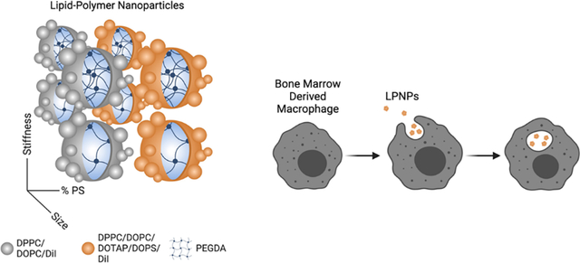
In this work, we created an array of eight lipid-polymer nanoparticles (LPNPs) that are small/large, soft/stiff, and with/without PS. These LPNPs consist of a polymer core to impart physical attributes surrounded by a lipid coating to aid in cellular interactions. When given to bone marrow-derived macrophages, LPNPs that were large, stiff, and contained phosphatidylserine (PS) were taken up significantly more than any other formulation, demonstrating the importance of all three physicochemical characteristics on endocytosis and indicating that these characteristics work better together than alone.

Footer

107 W Dean Keeton St. Austin, TX 78712

Google Maps

Email: suggs@utexas.edu Phone: (512) 232-1671

Fax: (512) 471-0616

Website by Kai Suggs

UT Home | Emergency Information | Site Policies | Web Accessibility | Web Privacy | Adobe Reader

© The University of Texas at Austin 2025