Longitudinal studies of antibody-mediated immunity have revealed that humoral immunity is often short-lived and appears to lack durability (Brouwer et a.l. 2020; Long et al. 2020). Thus far, much of the understanding of the immune response to COVID-19 has largely focused on peripheral blood samples from COVID-patients with diverse disease severity. Kanoko et.al investigated the alterations in secondary lymphoid organs, where adaptive immune responses are generated, to better understand the mechanisms behind humoral immune response to SARS-CoV-2. Because COVID-19 most significantly affects the lungs, architecture and lymphocyte populations of thoracic lymph nodes from patients were imaged and quantitatively analyzed. Spleen and peripheral blood samples were also examined.
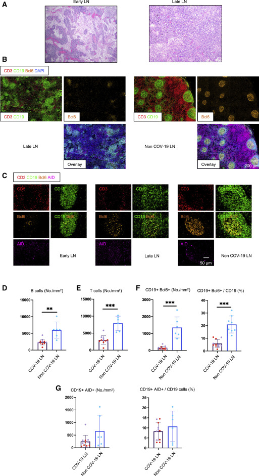
Germinal centers are important sites of selection of high-affinity B cells with long lifespans and pathogen-specific antibodies. Utilizing imaging approaches that preserve the architecture of human tissue, post-mortem lymph nodes were imaged from patients with severe disease who passed away within 8 days after admission and 15-36 days after admission (Figure 1A and 1B). In comparison to control lymph nodes from age-matched individuals, a dramatic loss of germinal centers and Bcl-6+ B cells was observed (Figure 1C and 1F). Bcl-6 is a critical transcription factor for T follicular helper cell differentiation and a key regulator of germinal centers.
Figure 1 – Early Loss of Germinal Centers and Bcl-6-Expressing B Cells in COVID-19 Thoracic Lymph Nodes (A) Hematoxylin-eosin staining of lymph nodes from early (left) and late (right) COVID-19 patients. (B) Low-power images of CD3 (red), CD19 (green), Bcl-6 (orange), and DAPI (blue) staining in a lymph node from a late COVID-19 patient (left) and a non-COVID-19 thoracic lymph node (right).(C) Representative multi-color immunofluorescence images of CD3 (red), CD19 (green), Bcl-6 (orange), and AID (purple) staining in lymph nodes from early (left) and late (middle) COVID-19 patients and a non-COVID-19 lymph node (right). (D and E) Absolute numbers of CD19+ B cells (D) and CD3+ T cells (E) in lymph nodes from COVID-19 patients (purple, n = 11) and non-COVID-19 patients (blue, n = 6). COVID-19 samples include early (purple, n = 5) and late (red, n = 6) COVID-19 patients. (F and G) Absolute numbers and relative proportion of Bcl6+ B cells (F) and AID+ B cells (G) in the pool of CD19+ B cells in lymph nodes from COVID-19 patients (purple, n = 11) and non-COVID-19 patients (blue, n = 6). COV-19, COVID-19; LN, lymph node. Mann-Whitney U test was used to calculate p value. Error bars represent mean ± SEM. **p < 0.01; ***p < 0.001. See also Figure S1 and Tables S1 and S2. (Kaneko et al. 2020)
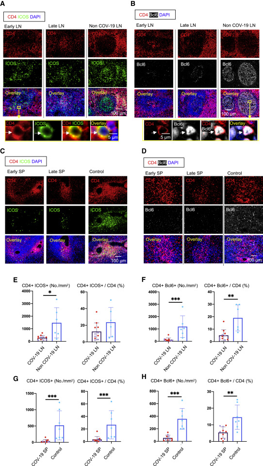
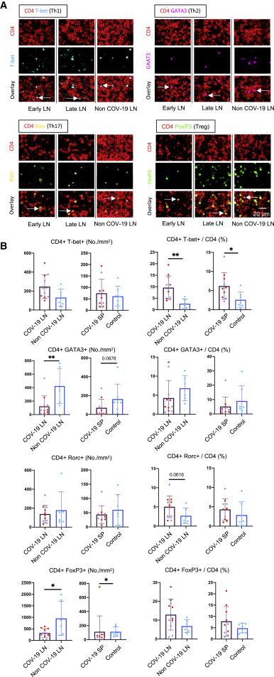
The authors also analyzed T follicular helper cell differentiation to better understand the possible mechanism behind germinal center loss. A striking loss of CD4+Bcl-6+ germinal center T follicular helper cells was found in spleen and lymph nodes (Figure 3B, 3D, 3F, and 3G). T follicular helper cells provide important signals in germinal centers for B cells to undergo clonal expansion and differentiate into memory B cells. The secondary lymphoid tissue was also examined for TNF-alpha expression, as germinal center loss in mouse models has been linked to an abundance of TNF-alpha and can be reversed with TNF-alpha blockade (Ryg-Cornejo et al, 2016; Popescu et al, 2019). TNF-alpha was found to be abundantly expressed, compared to activated lymphoid tonsil tissue held as a control, both inside and outside of the follicle. The authors hypothesize that the elevated synthesis of TNF-alpha, possibly induced downstream of Th1 activation which was also present at elevated levels, blocks the differentiation of T follicular cells (Figure 4).
Figure 3 – Loss of Germinal Center Type Bcl-6+ T Follicular Helper Cells in COVID-19 Lymph Nodes and Spleens (A) Representative multi-color immunofluorescence image of CD4 (red), ICOS (green), and DAPI (blue) staining in lymph nodes from early (left) and late (middle) COVID-19 patients and a non-COVID-19 control (right). Arrows indicate CD4+ ICOS+ TFH cells. (B) Representative multi-color immunofluorescence images of CD4 (red), Bcl-6 (white), and DAPI (blue) staining in lymph nodes from early (left) and late (middle) COVID-19 patients and a non-COVID-19 control (right). Arrows indicate CD4+ Bcl6+ GC-type TFH cells. (C) Representative multi-color immunofluorescence images of CD4 (red), ICOS (green), and DAPI (blue) staining in spleens from early (left) and late (middle) COVID-19 patients and a control (right). (D) Representative multi-color immunofluorescence images of CD4 (red), Bcl-6 (white), and DAPI (blue) staining in spleens from early (left) and late (middle) COVID-19 patients and a control (right). (E and F) Absolute numbers and relative proportions of CD4+ ICOS+ TFH cells (E) and CD4+ Bcl-6+ GC-type TFH cells (F) in lymph nodes from COVID-19 patients (purple, n = 11) and non-COVID-19 patients (blue, n = 6). COVID-19 samples include early (purple, n = 5) and late (red, n = 6) COVID-19 patients. (G and H) Relative proportions of CD4+ ICOS+ TFH cells (G) and CD4+ Bcl-6+ GC-type TFH (H) in spleens from COVID-19 patients (purple, n = 10) and controls (blue, n = 7). COVID-19 samples include early (purple, n = 4) and late (red, n = 6) COVID-19 patients. Mann-Whitney U test used to calculate p value. Error bars represent mean ± SEM. *p < 0.05; **p < 0.01; ***p < 0.001. See also Figures S2, S3, and S4 and Tables S1, S2, and S3. (Kaneko et al. 2020)
Figure 4 – Th1 Cells Are Expanded in Comparison to Other CD4+ T Cell Subsets in COVID-19 Thoracic Lymph Nodes and Spleens (A) Representative multi-color staining showing TH1, TH2, TH17, and T reg cells in lymph nodes form early (left) and late (middle) COVID-19 patients and a non-COVID-19 control (right; TH1: CD4+ [red] T-bet+ [light blue]; TH2: CD4+ [red] GATA3+ [purple]; TH17: CD4+ [red] RORg+ [yellow]; T reg cell: CD4+ [red] FOXP3+ [green]). (B) Absolute numbers and relative proportions of TH1, TH2, TH17, and T reg cells in lymph nodes and spleens (purple) from early (purple, lymph nodes: n = 5; spleens: n = 4) and late (red, n = 6) COVID-19 patients and controls (blue, lymph nodes: n = 6; spleens: n = 7). Mann-Whitney U test was used to calculate p value. Error bars represent mean ± SEM. *p < 0.05; **p < 0.01. See also Figures S5, S6, and S7 and Tables S1, S2, and S3. (Kaneko et al. 2020)The authors further analyzed preservation of AID-expressing B cells, non-germinal center derived B cells and circulating B cell population in patients with systemic inflammation, characteristic of severe COVID-19.
Overall, the findings in this paper contribute to understanding the mechanisms of disease progression in patients with COVID-19. Germinal centers in secondary lymphoid organs provide crucial microenvironments for the development of potent antibody responses and memory B cells; thus, the loss of GC responses at acute stages of infection, may account for the variable and often short-lived antibody responses observed in COVID-19 patients. This also draws light as to why expectations of natural herd immunity may be elusive.
References
Brouwer, Philip J. M. et al. 2020. “Potent Neutralizing Antibodies from COVID-19 Patients Define Multiple Targets of Vulnerability.” Science 369(6504): 643–50.
Long, Quan-Xin et al. 2020. “Antibody Responses to SARS-CoV-2 in Patients with COVID-19.” Nature Medicine 26(6): 845–48.
Kaneko, Naoki et al. 2020. “Loss of Bcl-6-Expressing T Follicular Helper Cells and Germinal Centers in COVID-19.” Cell 183(1): 143-157.e13.
Ryg-Cornejo, Victoria et al. 2016. “Severe Malaria Infections Impair Germinal Center Responses by Inhibiting T Follicular Helper Cell Differentiation.” Cell Reports 14(1): 68–81.Popescu, Maria, Berenice Cabrera-Martinez, and Gary M. Winslow. 2019. “TNF-α Contributes to Lymphoid Tissue Disorganization and Germinal Center B Cell Suppression during Intracellular Bacterial Infection.” The Journal of Immunology 203(9): 2415–24.
Leave a Reply